Endoscopy is a minimally invasive diagnostic procedure that examines the inner hollow organs. Endoscopy is performed using an optical instrument, which is called an endoscope. There are two types of endoscopes, rigid and flexible. Flexible endoscopes used in the review of the stomach, esophagus and colon (colonoscopy, Figure 1). Flexible endoscope owns flexible tip that allows good visualization of the stomach and intestines. Rigid Endoscope unlike no flexible flexible top, and so using it can not access certain parts of the body.
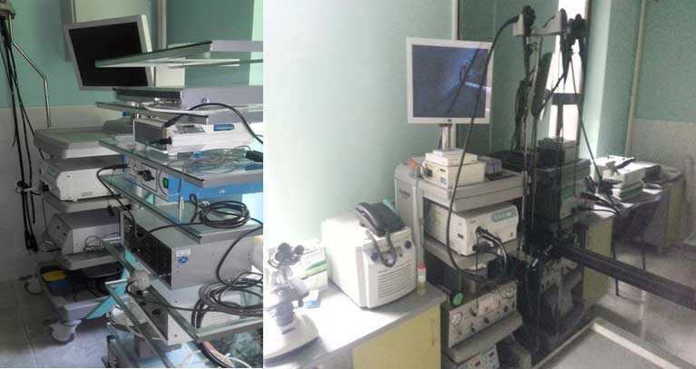
Figure 1. Video-endo-laparoscopic equipment
Flexible Endoscopy involves:
- Bronchoscopy (examination of the upper and lower airways, figure 2)
- Upper endoscopy or esophagus-gastro-duodenoscopy (esophagus, stomach and the initial part of the small intestine, Figure 3)
- Colonoscopy (examination of the large intestine – the colon, rectum and cecum, Figure 5)
Rigid endoscopy includes:
- Cystoscopy (examination of vaginal and urinary tract)
- Rectoscopy (examination of the rectum)
- Rhinoscopy (examination of the nasal cavity, Figure 4)
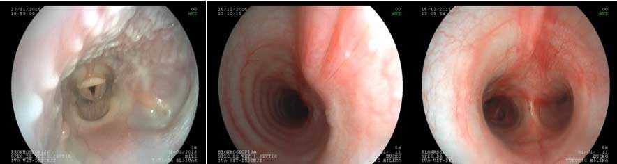
Figure 2. Bronchoscopy
Endoscopy is a complete procedure to get a complete visualization of the digestive system and front parts of the respiratory tract. In addition to visualization, endoscopy allows the taking of samples for biopsy of the stomach, intestines, nasal cavity, and also with the help of endoscope, it is possible to remove different foreign bodies from the esophagus and stomach.
Indications for bronchoscopy as chronic cough, wheezing and suspicion of the presence of bone bodies in the upper airways.
Indications for gastroscopy or an upper endoscopy chronic weight loss, blood in stools, frequent diarrhea, prolonged vomiting and presence of a foreign body in the esophagus or stomach.
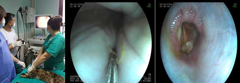
Figure 3. Gastroscopy
Indications for rhynoscopy bleeding from the nose, the constant presence of ooze from the nose, the presence of tumor in the nasal cavity and the presence of a foreign body.
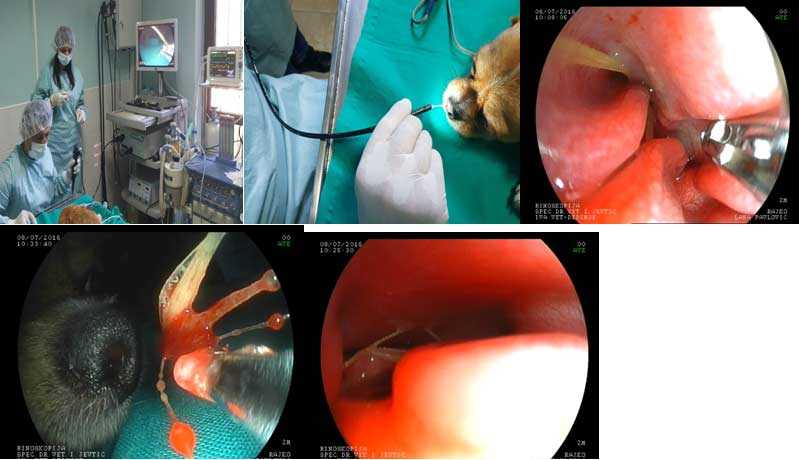
Figure 4. Rhynoscopy
Indications for cystoscopy were difficult urination and inability to control urination.
Endoscopy as a diagnostic procedure is performed under general anesthesia and prior to performing the procedure it is necessary to do blood tests. In addition to the findings it is necessary to prepare the patient for endoscopy, and it is reflected in the fact that the patient should be deprived of food for 12 to 24 hours and water 8 hours before the intervention.
Endoscopy procedures as an average lasts about 30 minutes and animals are lying on the left side.
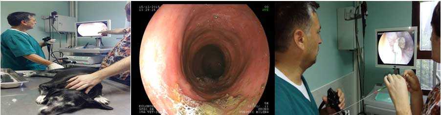
Figure 5. Colonoscopy
The use of stents, balloon catheters and gastrostomy catheter in dogs and cats
During upper endoscopy (rhinoscopy and traheoskopija) and lower (bronchoscopy) airway can be observed various disorders. During the examination of the upper airway most common problems that occur in dogs and cats are:
- Laryngeal paralysis,
- Brachiocephalic syndrome, and
- The collapse of the trachea
The collapse of the trachea (Figure 6.) is a problem that is most common in small breeds of dogs, primarily Yorkshire terriers, but usually characterized by the presence of chronic cough and shortness of breath.
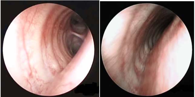
Figure 6. A. and B. The collapse of the trachea
The diagnosis can be set on the basis of radiography, fluoroscopy or traheoscopy. In our clinic definitive diagnosis is always by using endoscopic examination. Treatment depends on the severity of the clinical picture, other concomitant health problems, as well as places of localization of tracheal collapse. The treatment can be performed by cough medication, stress reduction, regulation of body weight, avoiding exerting pressure on the trachea leash for walking dogs, but usually we recommend the installation of tracheal stent thus solves the long-term health problem in your dog or cat (Figure 7).
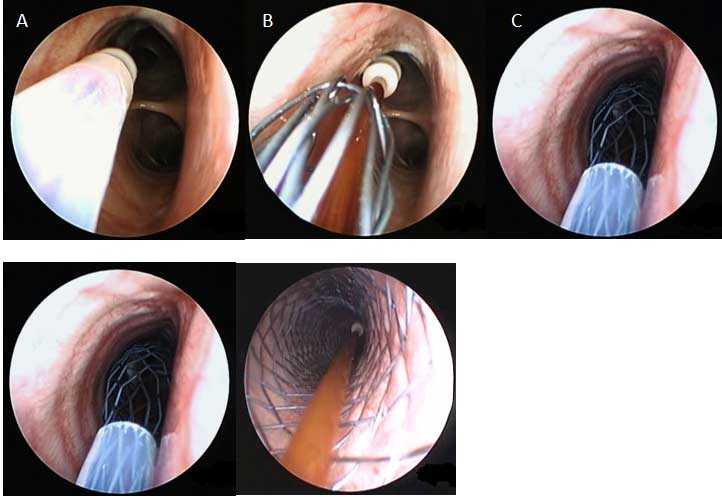
Figure 7. A, B, C, D and E Installation of tracheal stent
Installing the stent may also be made in other luminous bodies if there is a disorder that can not be otherwise disposed of. Thus, in the gastrointestinal tract when expelling gastroesophagoscopy stents are most often used in disorders in the esophagus (Figure 8). Most often used in order to palliation of inoperable malignant dysphagia, or can be used, although less frequently, and in the spread narrowed anastomoses, in covering the fistula and the expansion of benign stenosis.
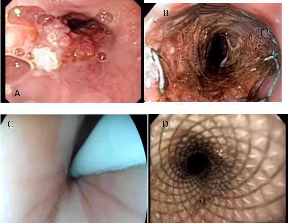
Figure 8. A. Complete obstruction of growing neo plastic in the esophagus B. Stenting as a method of treating a condition shown in Figure 8A.
C and D. Stricture of the esophagus and carotid stenting as a solution to this health disorder.
In cases where there is a stenosis of the esophagus and difficult passage of food through the esophagus to the stomach, in our clinic we perform endoscopic esophageal expansion, in places like this, using a special balloon catheter. It performs dilation of the esophagus by minimally invasive method and thus eliminates the problem. If the problem can not be solved using balloon dilatation, we recommend installing exophagial stent. These procedures are performed in dogs and cats.
In such a situation where we can not use the balloon dilatation and stent as a solution for the problem of passage of food through the esophagus, then in dogs and cats we do gastrostomy with special catheters where we endoscopic introduce them into the stomach and set so that the food is inserted by the owners from the outside directly into the stomach. This method is minimally invasive, performed under general anesthesia and for the pet it is the only way to overcome the problems in the esophagus (Figure 9).
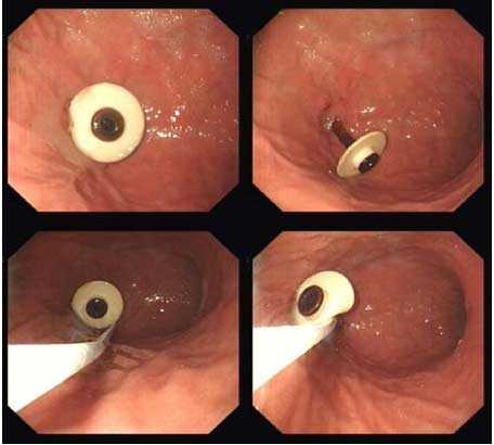
Figure 9. Gastrostomy