Laparoscopic surgery is a minimally invasive surgery, a technique that allows the intervention to be performed by using multiple small abdominal incisions. Specialized camera with fiber-optical fibers (laparoscope) is introduced through one of these portals in order to allow visualization of the internal contents of the abdomen. Similarly, through other portals surgical instruments necessary for the intervention are inserted into the abdominal cavity.
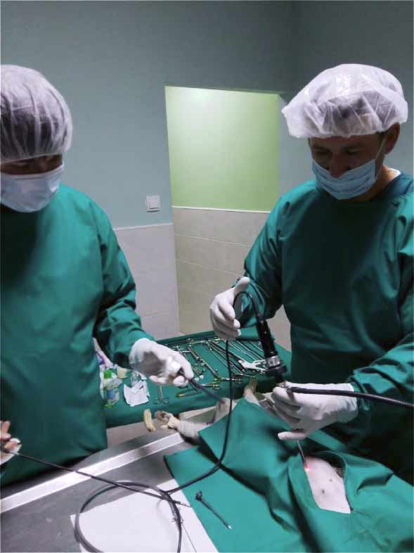
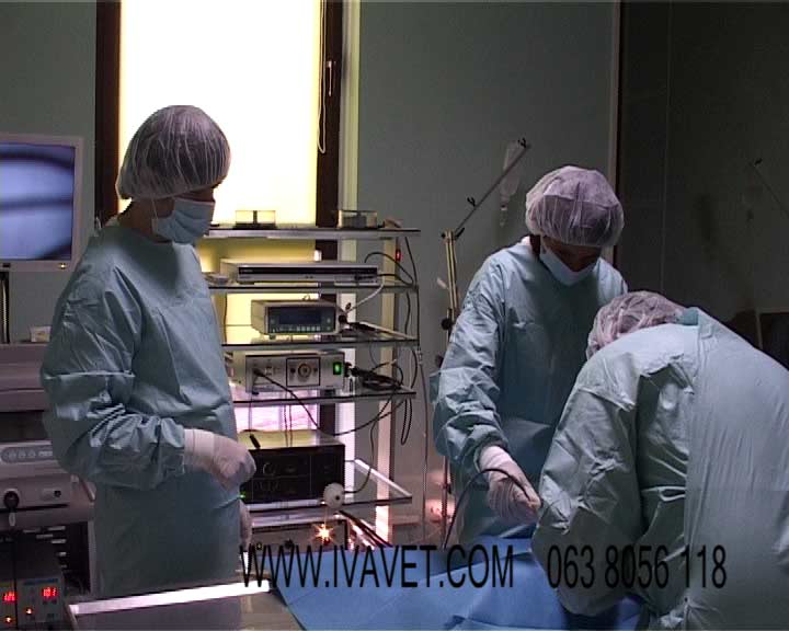
Laparoscopic surgery at IVA VET clinic is performed by the team of experts. IVA VET surgical team (picture 1.) utilizes advanced technology for the prophylactic, diagnostic, and therapeutic surgical procedures.
The most common type of surgery performed using minimally invasive technique is ovariectomy. This procedure is performed to prevent unwanted offspring, and to reduce the risk of infections and cancers of the female reproductive tract. Compared with traditional open ovariohysterectomy, laparoscopic ovariectomy is technically less complicated and timeconsuming.
Further, in a study published in the 2005 Journal of the Veterinary Medical Association has been documented that laparoscopic surgery diminishes pain, reduces the risk of hemorrhage and speeds recovery times up to 65%. In table 1. are presented laparoscopic surgery advantages over traditional open surgery.
Laparoscopic surgery advantages over traditional open surgery are numerous
| LAPAROSCOPIC SURGERY |
TRADITIONAL OPEN SURGERY |
| * Small incisions (less tissue trauma) | * Longer incision (extensive trauma) |
| * Quick recovery time-one day | * Long recovery time-up to two weeks |
| * Low risk of infection- in proportion with size of surgical wound | * High risk of infection – in proportion with size of surgical wound |
| * No formation of postsurgical hernia and adhesions |
* Possible formation of postsurgical hernia and adhesions |
| *The intestines (bowel movements) are usually somewhat lazy, most commonly for a day | * The intestines (bowel movements) are usually somewhat lazy for a few days |
| * Significantly smaller scar tissue after surgery | * Highly visible and characteristic scars |
| * No need for Elizabethan collar or body | * Mandatory use of E-collar or body |
| * No tearing or need to remove surgical thread | * Possible risk of tearing thread |
| * Less stressful postsurgical recovery | * More stressful postsurgical recovery |
| * Reduced risk of bleeding during and after surgery |
* More frequent appearance of bleeding during and after surgery |
| * Less use of painkillers | * Required use of painkillers |
 |
 |
| Picture A- Laparoscopic surgery | Picture B- Traditional open surgery |
The most common reasons for laparoscopic intervention are:
– Diseases causing acute or chronical pain in abdominal or pelvic cavity.
– Visualization of miscellaneous growths and patches in abdominal cavity, and collection of various samples (biopsy) for pathohistological examination.
– Ovariectomy and ovariohysterectomy
– Determining possible causes for free fluid accumulation in abdomen.
– Cancer staging for specific tumors.
– Surgical removal of tumors or organ invaded by tumor.
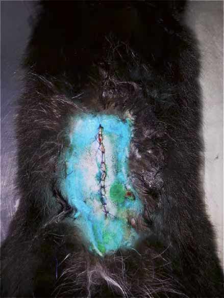
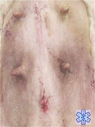
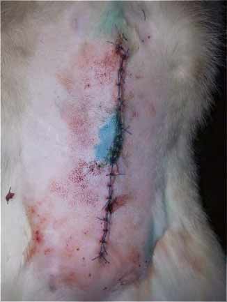
Picture 2. On left is presented tissue appearance after laparoscopically performed surgery. On right is presented tissue appearance after traditional open surgery.
Laparoscopic procedures in abdomen cavity:
– Ovariohysterectomy (in this procedure both, ovaries and uterus are removed)
– Ovariectomy (spay), only the ovaries are removed
– Sterilization of male dog
– Cancer and cystic kidney surgery
– Hernia Repair
– Ultrasound guided percutaneous sampling (biopsy) of abdominal organs
– Surgery of polycystic ovaries
– Gastropexy (Bloat/GDV Prevention)
– Removal of various tumor masses
Pre-operative assessment
Animal owners should expect the following procedures to be preformed during the preparation for the laparoscopic intervention:
1. General physical examination to determine animal health status.
2. Laboratory blood analysis (1.Blood chemistry panel—Used to evaluate organ function, electrolyte status, hormone levels, and more; 2.Complete blood count—Gives us information on hydration status, anemia, infection, clotting ability, and the ability of the immune system to respond to disease)
3. Laboratory urine analysis (Checks the condition of the urinary and genital tracts and screens for conditions such as diaďetes, liver disease, and Cushing͛’s disease)
4. Abdominal ultrasound (enabling a partial examination of the abdominal cavity- A noninvasive, real-time, moving picture of your pet͛’s abdomen, chest and heart)
Contraindication for Laparoscopic surgery
Absolute contraindications
– Diaphragmatic hernia
– Septic peritonitis
– Conditions in which conventional surgical intervention is obviously indicated
Relative contraindications
– Obesity (obscure the view of many organs)
– Poor patient condition
– Ascites
– Poor clotting time
– Patient body weight <2 kg (instrument size)
– Patient that is a poor anesthetic risk or an extreme surgical risk
Patient preparation before surgery
Owners should withhold food for 6-12 hours (over night) before surgery.
Anesthesia for laparoscopic surgery
Laparoscopic surgery is routinely performed in general anesthesia.
Laparoscopic Surgery Procedures in general
Preoperative preparation of patient
Empty urinary bladder for a better visualization of the abdominal cavity and to minimize the danger of tapping. Position the patient. Aseptically prepare the surgical field in the standard fashion.
Surgery
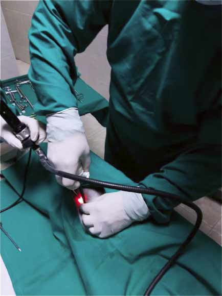
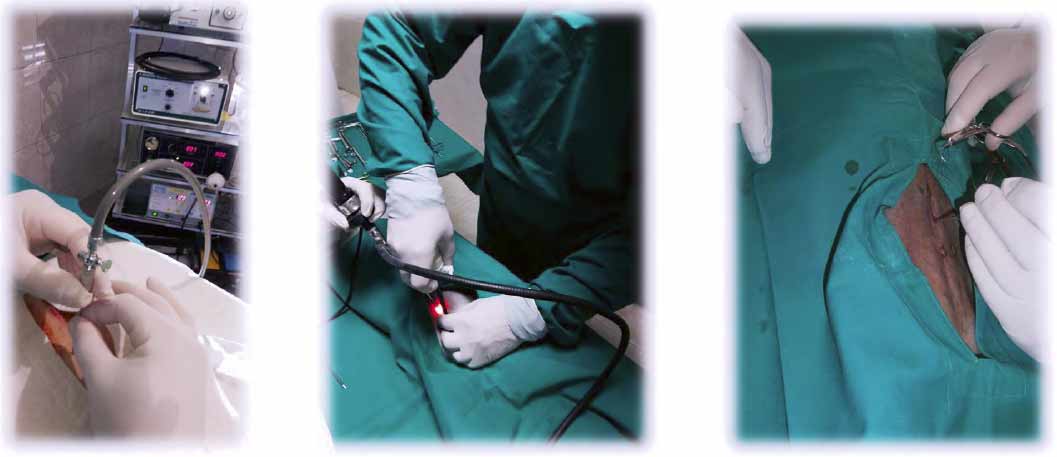
A surgeon makes one initial incision (picture 3.) commonly in the navel area. Then, a small needle is inserted through this incision, through which carbon dioxide gas can be pumped into the abdomen to inflate it allowing for better visualization of the abdomen’s contents.
Pressure in abdomen (picture 4.) must not be higher than 15 mm Hg (maintain the abdominal insufflation pressure at 12 to 15 mm Hg). If pressure in abdomen is higher, patient respiration will be impeded. Next, a laparoscope is inserted through one of the incisions. The camera illuminates the interior of the abdomen and transmits high-quality, magnified images to a video screen in the operating room, allowing for precise maneuvering. After that, surgeon can begin with organ examination. If required, more incisions are made on abdomen to insert instruments (basic equipment and instruments required to perform laparoscopic surgery in dogs and cats are listed in table 2.), and perform the surgery or/and sample collections (biopsy). Once the procedure is completed, the carbon dioxide is let out of the abdomen and the incisions are closed using stitches or clips.
Table 2. Basic equipment and instruments
1.Endoscopic tower monitor – camera system – light source – C02-insufflator – recording system – suction device
2. Laparoscope
3.Laparoscopic staplers
4.Retractors
5. Blunt and port trocar
6. Veress Needle
7. Suture material
8.Bipolar forceps (for coagulation)
9.Grasping forceps
10. Scissor forceps
Picture 4
 |
 |
| Picture A – Laparoscopic surgery | Picture B – Traditional open surgery |
Postoperative procedures
Any collected tissue or liquid sample during laparoscopic surgery will be sent for further pathohistological examination. Results of those analyses can be expected few days after the procedure.
Postoperative recovery after the laparoscopic surgery is much faster, safer and less stressful for your animal companion.
Duration of laparoscopic surgery
Depending on the complexity of procedure, laparoscopic surgery can last anywhere from half an hour to several hours.